Why Drosophila?
Why flies?
Start with the animal. Keep moving inward.
01Adult female
Drosophila lets us follow a biological question across scales—from an adult female to the cells at the beginning of an ovariole.
02Inside the abdomen
The exterior is only the first layer.
Inside the abdomen, paired ovaries bring repeated developmental structures into one compact system.
The faded exterior remains as orientation while the model reveals the ovaries beneath it.
03Repeated structure
An ovary is built from repeated ovarioles.
Each ovariole places early development at its anterior tip and progressively older egg chambers toward the posterior.
The model shows a representative arrangement. Its generated ovariole count is not a universal invariant.
04Anterior tip
At the anterior tip, development begins.
The germarium organizes germline cyst development among surrounding somatic cells.
Escort cells support the anterior region; follicle stem cells and early follicle cells occupy the transition into the first egg chambers.
How these stem cells renew tissue05Stem-cell community
A stem-cell population builds the follicular epithelium.
Follicle stem cells lie along the germarium’s outer surface, between escort cells and early follicle cells.
This simplified anatomy follows the Kalderon Lab model: a community of stem cells supplies the follicle cells that surround developing egg chambers.
See the lineage-tracing experiments06BioVision data
The model is a map. The lab works with real tissue.
The teaching model on the right is representative anatomy; it is separate from the source-paired BioVision reconstruction in the player.
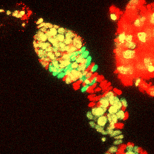
Microscopy paired with its BioVision reconstruction. The meshes are tracked segmented regions; source contour colors can repeat and do not identify cell type.
About this dataset
Source-paired BioVision reconstruction and lossless maximum-intensity microscopy projection at 31 biological timepoints, sampled every 531 seconds (8.85 min).
The orthographic 3D view shares the microscopy field. Biological time advances through the paired timepoints while camera rotation, pan, and zoom remain independent. Frame-to-frame movement is shown without artificial stabilization.
Model qualification
Read the model at the right resolution.
Scientifically informed adult-female external teaching model, not specimen-exact or morphometrically validated
Detailed limitations
- The model is procedural rather than a micro-CT, photogrammetry, or direct specimen scan.
- Dimensions, spacing, identity colors, transparency, authored ink, and lighting are illustrative and support explanation rather than measurement or biological optics.
- Cell-level detail is concentrated in one left ovariole; non-focus ovarioles and the posterior reproductive context are simplified.
- The teaching model does not represent every follicular cell, and generated ovariole counts are representative rather than invariant.
- The follicle stem-cell population is a representative static arrangement based on the Kalderon Lab model, rather than a fixed cell census.
Sources and scope
- Kalderon Lab — Adult Stem Cell Research
Research overview and the approximate follicle stem-cell population described by the lab.
- Dong et al. (2023), BMC Biology
Germarium organization, escort-cell context, and germline development.
- Melamed et al. (2023), PLOS Genetics
Spatial organization, behavior, and differentiation in the follicle stem-cell community.
- Reilein et al. (2017), Nature Cell Biology
A dynamic follicle stem-cell population maintained by spatially patterned signals.
Continue into the research