Find research sections and publications. Use arrow keys to select a result, Enter to open it, and Escape to close search.
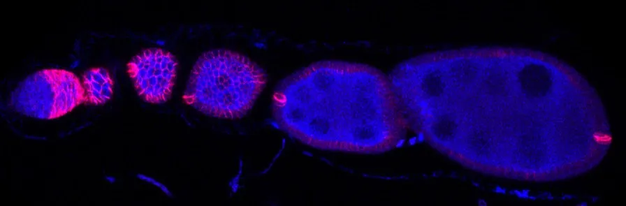
Figure 01
Egg development proceeds along an ovariole. DAPI marks nuclei in blue; Fas3, shown in red, marks the follicle-cell epithelium.
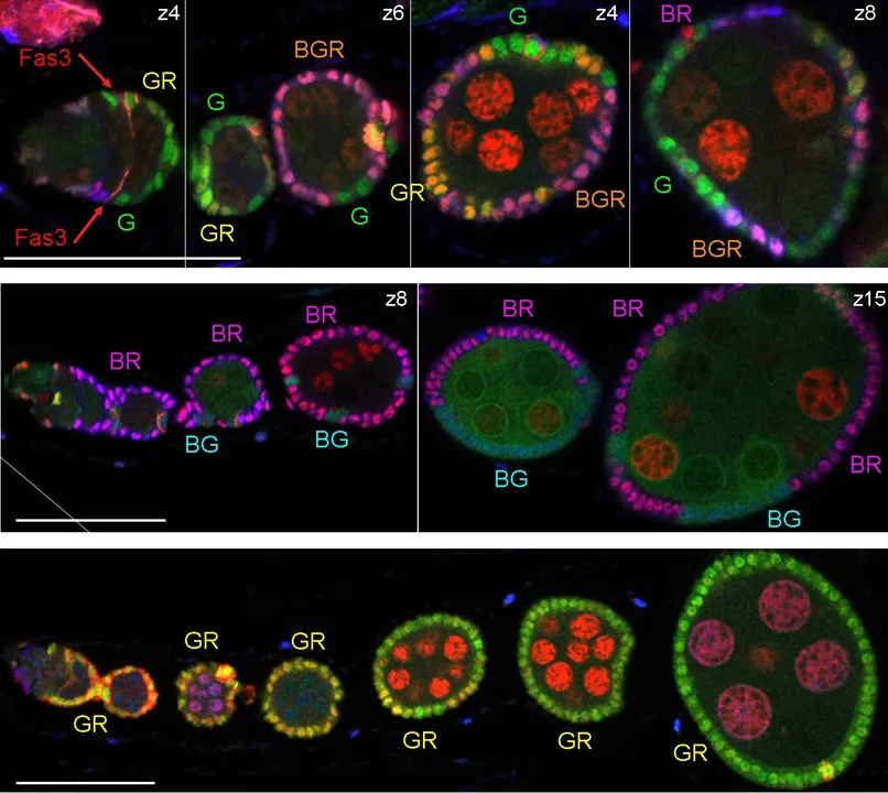
Figure 02
The six-color labeling system distinguishes descendants of labeled FSCs. Rows show separate samples after label induction, with the original labels and scale bars retained.
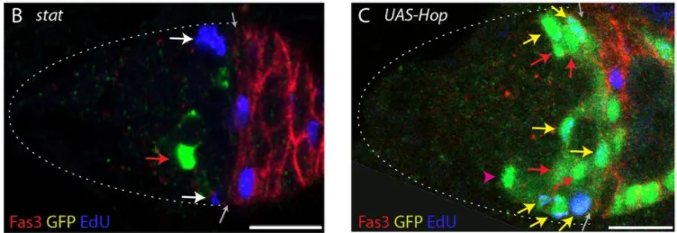
Figure 03
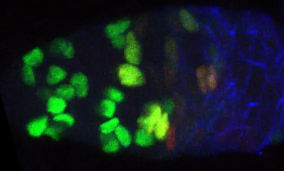
Removing STAT reduces proliferation and lineage retention. Elevated Hop, a JAK kinase, increases proliferation and the number of marked FSCs. EdU is blue in this figure.
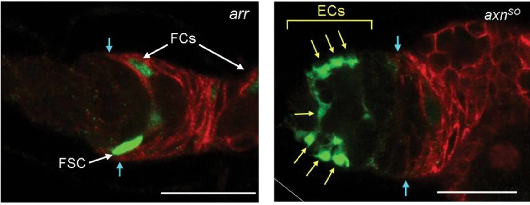
Figure 04
Loss of Wnt reception through arr shifts marked cells posteriorly and prevents escort-cell production; elevated signaling in axn mutants favors anterior positions and escort cells.
Figure 05
Differential E2F-GFP and CycB-RFP signals help distinguish cell-cycle phases in follicle stem cells and escort cells.
Figure 06
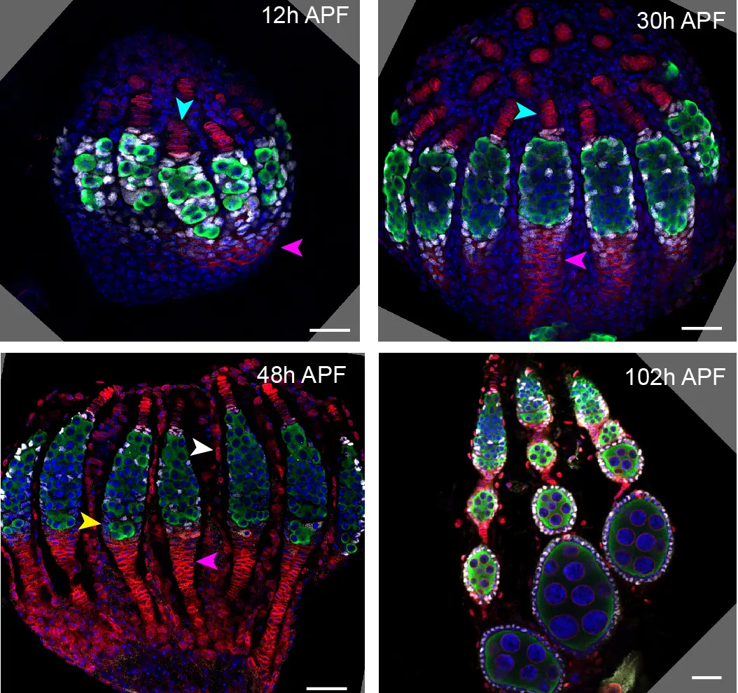
Vasa marks germline cells in green; Traffic Jam marks somatic precursors in white; DAPI labels nuclei blue. These are separate developmental samples.
Figure 07
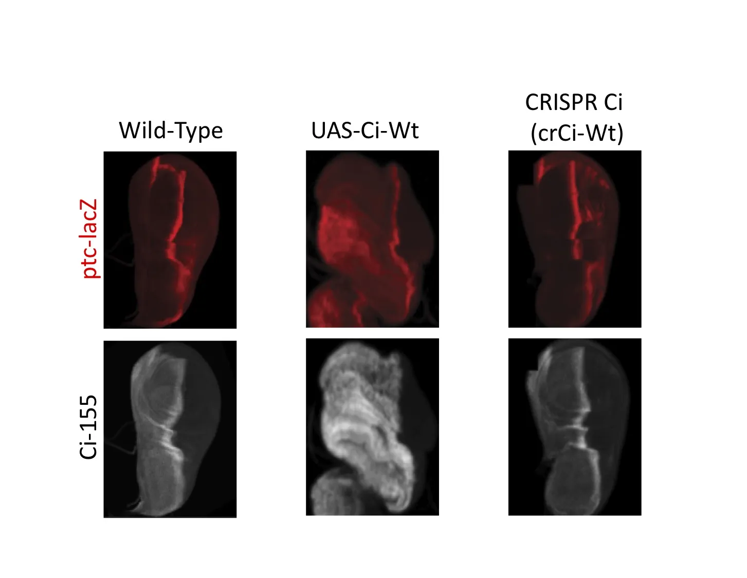
In these examples, UAS-Ci produces excess Ci and altered disc shape. Ci expressed from the edited endogenous locus more closely resembles wild type.
Figure 08
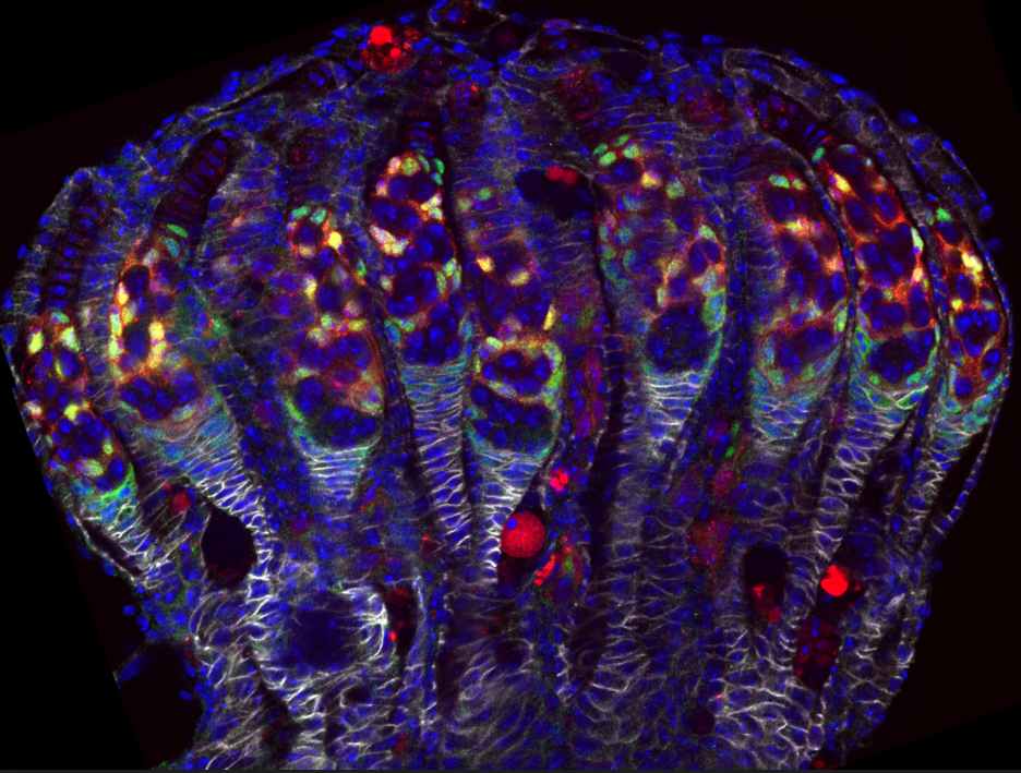
Image of a pupal ovary composed of multiple ovarioles with a domain of Wnt signaling shown by the expression of Frizzled3-RFP in red, and precursors to Escort cells and Follicle Stem Cells shown by Traffic Jam staining in green.